Biochemical test is a test that identifies the different types of microorganisms. Some biochemical tests uses the presence of catalase and other different waste products to identify different types of microorganisms while other tests isolate specific groups of microorganisms by inhibiting growth of other bacteria to identify the bacteria. Some tests even use different staining techniques to identify different microorganisms.
There are many Biochemical Test done to identify the the different microorganisms. We have chosen a few experiments to present in our webpage. To view our topic, refer to the "Topic" section on the side box.
Aseptic broth transfer and colony selection
Aseptic broth transfer and colony selection
What is it about?
Aseptic broth transfer and colony selection is all about performing basic bacteriological transfer techniques using broth and agar cultures. This will also determine how well one handles bacteriological cultures aseptically.
Procedures:
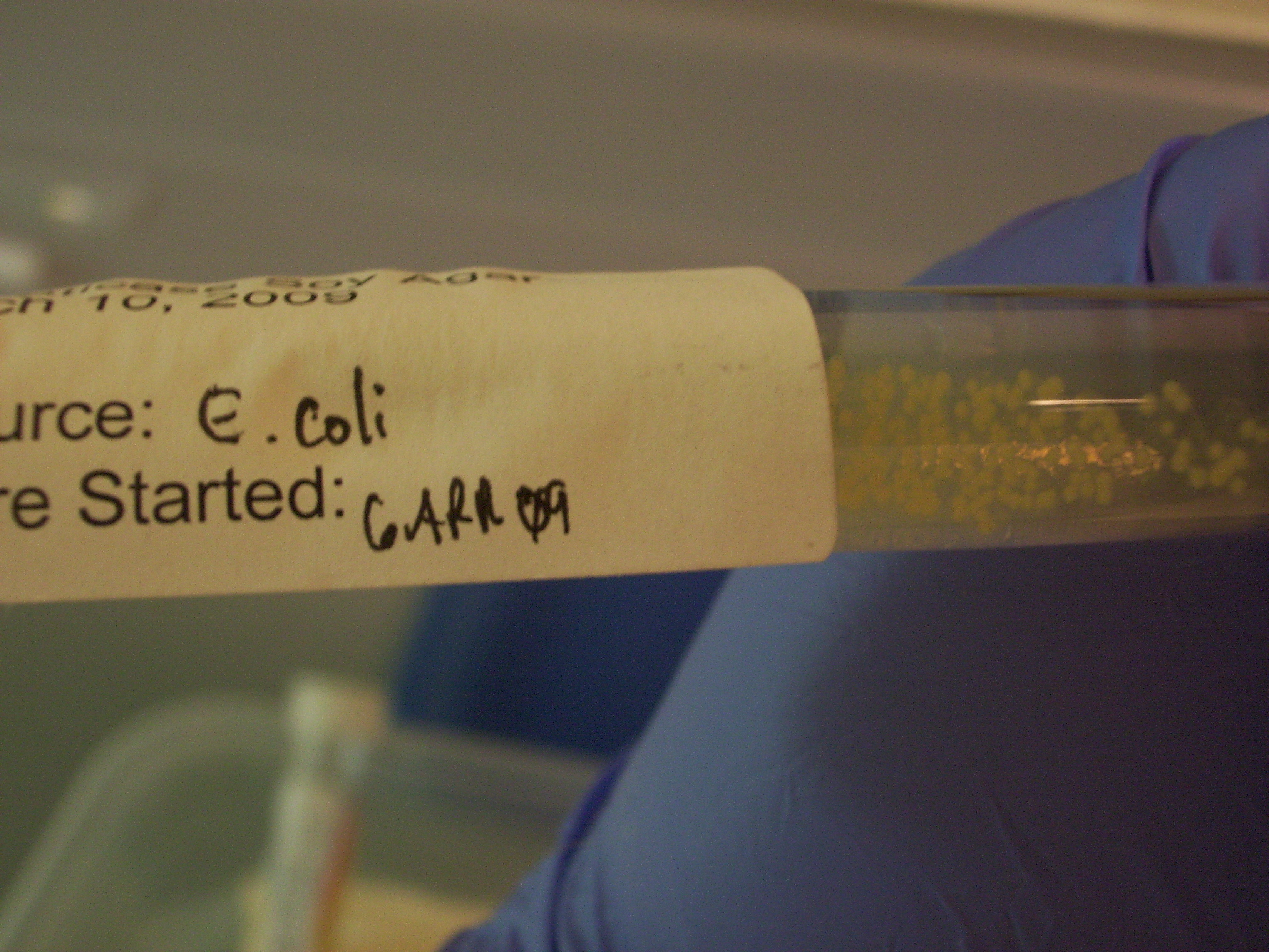
To start off, we have samples of Escherichia coli in nutrient broth, Micrococcus luteus on an agar slant, and Escherichia coli on a nutrient agar streak plate.
Firstly, we would use an inoculating loop to transfer E.coli from the broth culture into another test tube of fresh nutrient broth.
Next, we would use the inoculating loop to take a sample of M.luteus from the agar slant and transfer it into a test tube of fresh nutrient broth.
Then, we would use the inoculating loop to transfer a sample of E.coli from the nutrient broth and streak it onto a test tube of fresh agar slant.
Lastly, we would use the inoculating loop to take a sample of E.coli from the nutrient agar streak plate, and transfer into a test tube of fresh nutrient broth. (the inoculating loop is flame sterilized after each transfer)
Results:
E.coli from nutrient broth into nutrient broth
Broth turns turbid
Sediment is seen
M.luteus from agar slant to nutrient broth
Broth turns creamy yellow
Sediment is seen
E.coli from nutrient broth to agar slant
Pellicle is seen at the top of the tube
E.coli from nutrient agar streak plate into nutrient broth
Broth turns cloudy
Sediment is seen
However if any step has been carelessly done, it’ll result in…CONTAMINATION!
Gram Staining
GRAM STAINING
Microorganisms are not only very small, they are also transparent. Stains and dyes are used to color bacterial cells and increase their contrast so they can be seen more easily with a microscope. Dyes are organic compounds composed of a chromophore group, which gives the dye its color, and an auxochrome, which binds to the bacteria.
What is being tested?
A Gram stain is used to determine if bacteria are present in an area of the body that is normally sterile, such as spinal fluid. A sample from the infected area is smeared on a glass slide and allowed to dry. A series of stains and a decolorizer is applied. The stained slide is then examined under a microscope where bacteria appear either purple (gram positive) or pink (gram negative).
The test is named for Dr. Christian Gram, who invented the process. A Gram stain can predict the type of bacteria causing an infection, such as pneumococcal pneumonia or a staphylococcal abscess. Viruses cannot be seen with a Gram stain since they lack the cell wall, which takes up the stain.
Procedures for gram staining:
Place a slide with a bacterial smear on a staining rack.
Stain the slide with CRYSTAL VIOLET for 1 - 2 minutes.
Rinse off the stain with distilled water.
Flood slide with IODINE for 1 - 2 minute.
Rinse the slide with distilled water.
Add ALCOHOL decolourizer (Acetone) until dye no longer runs off from the smear.
Wash slide with distilled water to prevent additional decolourization.
Cover the smear with SAFRANIN for 2 minutes.
Rinse with distilled water.
Blot dry the slide.
So how does it work?
In Gram positive bacteria, the purple crystal violet stain is trapped by the layer of peptidoglycan which forms the outer layer of the cell.
In Gram-negative bacteria, the outer membrane prevents the stain from reaching the peptidoglycan layer in the periplasm. The outer membrane is then permeabilized by acetone treatment, and the pink safranin counterstain is trapped by the peptidoglycan layer.
Results of gram staining :
Gram Positive Bacteria
Gram Negative Bacteria
Selective n Differential Media
Selective and Differential Media
Selective and differential media are used to isolate or identify particular organisms.
Selective media allow certain types of organisms to grow, and inhibit the growth of other organisms. The selectivity is accomplished in several ways. For example, organisms that can utilize a given sugar are easily screened by making that sugar the only carbon source in the medium. On the other hand, selective inhibition of some types of microorganisms can be achieved by adding dyes, antibiotics, salts or specific inhibitors which affect the metabolism or enzyme systems of the organisms.
For example, media containing potassium tellurite, sodium azide or thallium acetate (at concentrations of 0.1 - 0.5 g/l) will inhibit the growth of Gram-negative bacteria. Media supplemented with penicillin (5-50 units/ml) or crystal violet (2 mg/l) will inhibit the growth of Gram-positive bacteria. Tellurite agar, therefore, is used to select for Gram-positive organisms, and nutrient agar supplemented with penicillin can be used to select for Gram negative organisms.
Differential media are used to differentiate closely related organisms or groups of organisms. Owing to the presence of certain dyes or chemicals in the media, the organisms will produce characteristic changes or growth patterns that are used for identification or differentiation. A variety of selective and differential media are used in medical, diagnostic and water pollution laboratories, and in food and dairy laboratories.
MANNITOL SALT AGAR (MSA)
Mannitol salt agar is a selective medium used for the isolation of pathogenic staphylococci. The medium contains mannitol, a phenol red indicator, and 7.5% sodium chloride. The high salt concentration inhibits the growth of most bacteria other than staphylococci. On MSA, pathogenic Staphylococcus aureus produces small colonies surrounded by yellow zones. The reason for this change in color is that S. aureus ferments the mannitol, producing an acid, which, in turn, changes the indicator from red to yellow. The growth of other types of bacteria is generally inhibited.
MacCONKEY'S AGAR
MacConkey’s agar is a selective &differential plating medium designed to grow Gram Negative bacteria and stain them for lactose fermentation. It contains bile salts (to inhibit most Gram-positive bacteria, except Enterococcus and some species of Staphylococcus), crystal violet dye (which also inhibits certain Gram-positive bacteria), neutral red dye (which stains microbes fermenting lactose), lactose and peptone. The growth of Gram-positive organisms is inhibited because of the crystal violet and bile salts in the medium. Lactose is the differential component of McConkey Agar. The bacteria that can ferment lactose changes the pH of the media to become acidic and the media will turn pink due a pH indicator in the agar. Bacteria that does not ferment lactose will grow on the plate without casing a colour change as no acid was produced.
HEKTOEN-ENTERIC AGAR (HE)
HE agar is a selective & differential agar primarily used to recover Salmonella and Shigella from patient specimens. HE agar contains 3 carbohydrate (lactose, salicin and sucrose) and two indicator dyes (Bromthymol Blue and Acid Fuchsin). The high bile salt concentration inhibits growth of all gram-positive bacteria and retards the growth of many strains of coliforms. Acids may be produced from the carbohydrates, and acid fuchsin reacting with thymol blue produces a yellow colour when the pH is lowered. Sodium thiosulfate is a sulphur source, and H2S gas is detected by ferric ammonium citrate. Rapid lactose fermenters (e.g. E.coli) are moderately inhibited and produce bright orange to salmon pink colonies. Salmonella colonies are blue-green, typically with black centres from H2S gas. Shigella appears greener than Salmonella, with the colour fading to the periphery of the colony. Proteus strains are somewhat inhibited, forming small transparent and more glistening or watery in appearance than species of Salmonella or Shigella.
BLOOD AGAR
Contains mammalian blood (usually sheep or horse), typically at a concentration of 5–10%. BAP are an enriched, differential media used to isolate fastidious organisms and detect hemolytic activity. β-hemolytic activity will show complete lysis of red blood cells surrounding colony. Examples include Streptococcus haemolyticus. α-hemolysis will only partially lyse hemoglobin and will appear green. An example of this would be Streptococcus viridans. γ-hemolysis (or non-hemolytic) is the term referring to a lack of hemolytic activity.
6 different bacteria ( Escherichia coli, Staphylococcus aureus, Entrobacter aeurogenes, Streptococcus faecalis, Proteus vulgaris and Micrococcus luteus ) .
3 plates each of Blood agar, MacCokey agar, MSC agar and HE agar.
Inoculating loops.
Marker pen.
Procedures:
Divide each of the agar plates into half using the marker pen.
Streak each bacteria on half of each four plates.
Close the lid and inverse the petri dish.
Incubate at 37°C for 48 hours and store at 4°C after 48 hours.
Observe the plates and combine the results for all six microorganism on each plate.
Results:
(click to view results)
Blood Agar
MacConkey Agar
MSA
HE Agar
Endospore Staining
Structral Staining for Endospores
Some Bacteria such as Bacillus and Clostridium have the ability to produce endospore. Endospores are resistant to heat, drying, radiation and various chemical disinfectants.
Endospores formation (sporulation) :
It occurs through a complex series of event. One endospore is produced within each vegetative bacterium. Once the endospore is formed, the vegetative portion of the bacterium is degraded and dormant endospore is released.
Endospore contains :
DNA, ribosome, cytoplasmic membrane, cell wall and then everything else that are essential for bacterial life.
These cell constituents are then surrounded by a series of protective coats including a cortex, a spore coat, and sometimes an exosporium. The dehydrated state of the endospore also contributes to its resistance.
Endospores are able to survive for a long period of time until environmental conditions become favourable for growth. The endospore then germinates producing a single vegetative bacterium.
Endospores are difficult to stain, due to its resistant nature to heat and chemicals. Strong dyes and vigorous staining conditions are required. However once stained, endospores are hard to decolorize. Since few bacterial genera produce endospores, the endospore stain is good diagnostic test for species of Bacillus and Clostridium.
Procedure :
Smear preparation and heat fixing of smear.
Place a small piece of paper towel square over the smear and saturate with MALACHITE GREEN for 30-60 seconds.
Heat gently by passing the Bunsen burner under the slide for 5 minutes. Heat the slide until steam can be seen rising from the surface. Remove slide the heat until steaming stops; then gently reheat. As the dye evaporate, continually add more.
Allow slide to cool and wash the excess stain. Remove the paper towel square off the slide with a forceps and wash off excess stain with didtilled water.
Blot the slide dry and counterstain for 1 minute with 0.5% SAFRANIN.
Wash off the excess safranin with water, blot dry, and observe using oilimmersion microscopy.
Results :
The vegetative cells will appear red and the spores will appear green.
Flagella ; Motility Test & Hanging Drop Preparation
Flagella & Motality Test
Flagella
Many bacteria are capable of mortality due to the presence of specialised structures called flagella. The shape of the flagella about the bacterium is used in classification and identification.
The following flagella arrangements may be found.
Athrichous - no flagella present.
Monotrichous - a single flagellum at one pole.
Amphitrichous - single flagella at both poles.
Lophotrichous - two or more flagella at one or both poles of the cell.
Peritrichous - completely surrounded by flagella.
Motility Test -
This test is used to determine if organisms are motile by means of flagellum. The location of the flagella is determined by the bacterial species. Non-motile bacteria do not possess flagellum. Bacterial motility must be distinguished from Brownian motion. Feebly motile bacteria may require prolonged observation of individual cells. Motility results are difficult to determine for anaerobic bacteria. Only a positive result is significant. Some bacteria are motile at one temperature and non motile when incubated at another. Some bacteria become less motile in old cultures.
Materials :
Staphylococcus aureus ans Proteus vulgaris culture.
Inoculation needle.
Motility Test Agar
Procedures :
Inoculate the motility test agar with 1 of the bacteria listed above.
Use a straight inoculation needle.
Employing aseptic technique, stab down into the centre of the agar to about one-half of the way down.
Withdraw needle carefully along the same stab line.
Repeat with the other bacteria listed. Incubate at 37°C for 24 hrs. Label this tube.
Results :
Non-motile Organisms have bacterial growth along the line of the stab.
Motile Organisms have spreading or diffused bacterial growth from the stab line.
Hanging Drop Preparations -
Procedure :
Using sterile technique, transfer a loopful of culture to the centre of a clean glass cover slip.
Roll out 2 plasticine strips (ensure that they are not thick and about the length of coverslip.
Gently press a slide against the coverslip, turn it over quickly, leaving the drop suspended in the middle of the cover slip.
Examine the culture under high power objectives. Close the diaphragm opening, because the bacteria are unstained and they will be difficult to locate.
Focus on the edge of the droplet before attempting to locate individual cells. After the bacterial cells have been located, study them carefully for evidence of motion.
Results :
An example of a hanging drop slide
Catalase Test
Catalase Test
The catalase test is a biochemical test used by microbiologists to identify species of bacteria (whether they possess catalase). It requires the adding of a few drops of 3% hydrogen peroxide directly to the young broth culture or colonies on agar surface or clumps of cells on glass surfaces to determine the presence of catalase in the organisms. Catalase is a common enzyme found in nearly all living organisms which are exposed to oxygen (Aerobic organisms). Obligate Anaerobes do not have catalase as they do not use oxygen in their respiration.
Catalase catalyses the decomposition of hydrogen peroxide to water and oxygen according to the equations:
O2- → O2 + 2H2O
Superoxide dismutase
2H2O2 →2H2O + O2
Catalase Test -
Materials :
24 hours trypticase soy agar plate of Staphylococcus aureus and Streptococcus faecalis.
3% hydrogen peroxide solution.
Droppers
Procedures :
Add 2 to 3 drops of 3% hydrogen peroxide solution onto isolated bacterial colonies.
Observe and look out for any effervescences produced.
Results :
A Positive test is indicated by the production of gas bubbles or the presence of effevescence. Staphylococci and micrococcus are examples of genus of catalase-positive bacteria.
The white bubbles are oxygen gas produced from hydrogen peroxide.
The catalase test is Negative if there is no effervascence formed. Streptococci and enterococci are examples of genus of catalase-negative bacteria.
Streptococcus species is an example of aerobic bacteria that do not possess catalase. Catalase has also been observed in some anaerobic microorganisms, such as Methanosarcina barkeri. Thus, not all aerobes possess catalase and anaerobes do not possess catalase.
IMViC tests
IMViC Tests
The IMViC tests involve a set of tests which is used to differentiate bacteria from one another. For example, to differentiate organisms such as Klebsiella,Entezrobacter, and Escherichia coli.
There are mainly four individual test- the Indole test, methyl red test, Voges-Proskauer reaction and the citrate test.
Indole test
The purpose of the Indole test is to differentiate species of the family Enterobacteriaceae by testing the bacteria’s ability to produce Indole from amino acid tryptophan. When indole is produced, it reacts with Kovac’s reactant to create a cherry red-coloured layer.
Procedure:
1. Add 5 drops of Kovac’s reactant to each test tube of Trypticase soy broth culture.
2. If a cherry red-coloured layer is form, it shows that an Indole positive bacteria is present (eg. E.coli) as the bacteria produces an enzyme which cleaves tryptophan to produce indole and other products.
The Methyl Red and Voges-Proskauer Tests
The two tests are used to help differentiate bacteria species of the family Enterobacteriaceae. The methyl red test tests for acid end products from glucose fermentation whereas the Voges-Proskauer tests for acetoin production from glucose fermentation.
Procedure for methyl red test:
1. Add 4-5 drops of the indicator, methyl red to a test tube containing the bacteria culture.
2. If a red precipitate is formed, it is a positive test where glucose is being fermented to form acid, hence causing the pH to drop and form a red coloration. On the other hand, if a yellow precipitate is formed, it is a negative test.
Left- Negative test; Right – Positive test
Procedure for Voges-Proskauer Test:
1.Add 5 drops of the alpha-naphthol solution to a test tube containing the bacteria culture.
2. Add 5 drops of potassium hydroxide-creatine solution to the test tube and shake well for 1 minute.
3. If a red precipitate is formed, it is a positive test. Otherwise, there is no colour change which signifies a negative test.
[Positive test => acetoin produced; Negative test => acetoin not being produced]
Citrate test
This test helps to differentiate species of the family Enterobacteriaceae. It is selective for bacteria which is able to consume citrate as the only source of carbon and ammonium as its sole nitrogen source.
Procedure:
1. Inoculate bacteria onto the slant agar containing Simmon's citrate media.
Left- Positive; Right- Negative
When it is a positive test, the green Simmon’s Citrate Agar (pH of 6.9) will turn blue as the bacteria metabolizes citrate to produce an acidic product.
oh hello stranger
Hello we warmly welcome you to this website!
Proudly done by Gurinder, Joash, Marisa, Wahidah, Wei Jian, Atikah of PS0903 :D
Now...
Sing along with the bacteria song!
[bacteria, bacteria]
Bacteria- Jonathan Coulton
Bacteria, Bacteria
Look, there's Bacteria
Bacteria, Bacteria
You might not see them, but they're there
Bacteria, Bacteria
Everything you touch
Bacteria, Bacteria
That's right, Salmonella Bacteria
But we have to watch out for bacteria
That can spoil our chicken
Bacteria practically everywhere
Everywhere you look
in the kitchen
inside the cooler
in the dining area
in the rest rooms
on our raw chicken
And like I said
Bacteria, Bacteria
Look, there's Bacteria
Bacteria, Bacteria
You might not see them, but they're there
Bacteria, Bacteria
Everything you touch
Bacteria, Bacteria
That's right, Salmonella Bacteria
Salmonella grows on raw chicken, especially old chicken
moist foods like our salads
staph bacteria on the left and strep bacteria on the right
Salmonella, sigillum, clostridium perfringens
If you didn't wash your hands, they would become breeding grounds for
Bacteria, Bacteria
Look, there's Bacteria
Bacteria, Bacteria
You might not see them, but they're there
Bacteria, Bacteria
Everything you touch
Bacteria, Bacteria
That's right, Salmonella Bacteria
Fever, cramps and fever
Dysentery
Fever, fe-fe-fe-fever
Vomiting, vomiting
Chills
Cramps
Chills, and chills and cramps
One square inch
Half a billion Salmonella bacteria
These bacteria really sound serious
They are when they're left unchecked
And it could mean a trip to the hospital for someone
Our customers
wow
ourselves
Alright
our chicken
Alright
And our reputation
Alright, alright
You mean bacteria on me right now?
Clean, clean, and then clean again
Bacteria, Bacteria
Look, there's Bacteria
Bacteria, Bacteria
You might not see them, but they're there
Bacteria, Bacteria
Everything you touch
Bacteria, Bacteria
That's right, Salmonella Bacteria
Salmonella Bacteria
Salmonella Bacteria


